

Which Vertebrae Affect Which Nerves? A Spinal Nerve Guide
The short answer: A spinal nerve branches off at every level of the spine, and the level where it exits predicts the part of the body it serves. The neck (C1 to C8) runs the diaphragm, shoulders, arms, and hands; the mid-back (T1 to T12) runs the chest and abdominal wall; and the lower back and sacrum (L1 to S5) run the hips, legs, feet, bladder, and bowel. Match where your symptoms travel to the chart below and you can usually tell which level is involved.
When a nerve in your spine gets pinched, the pain, tingling, or weakness almost never stays where the problem is. A compressed nerve root in your neck can numb your thumb; one in your lower back can shoot pain to your big toe. That is because each spinal nerve exits at a specific vertebral level and travels to a specific territory of the body, so the symptom map is really a map of the spine. This guide walks the spine from top to bottom, shows which nerves leave at each level, lists the body parts and muscles they control, and explains the symptoms that show up when a nerve at that level is irritated, so you can connect what you feel to where it likely starts.
- The spine has 31 pairs of spinal nerves: 8 cervical, 12 thoracic, 5 lumbar, 5 sacral, and 1 coccygeal. A pair exits at every level.
- There are 8 cervical nerves but only 7 cervical vertebrae, so cervical nerves exit above their vertebra and C8 exits between C7 and T1; from T1 down, each nerve exits below its vertebra.
- Cervical levels drive the shoulders, arms, hands, and the diaphragm; thoracic levels wrap the chest and abdomen; lumbar and sacral levels drive the legs, feet, bladder, and bowel.
- Doctors use two body maps, dermatomes (skin sensation) and myotomes (muscle strength), to trace a symptom back to a single nerve root.
- Saddle numbness, new bladder or bowel changes, or weakness in both legs can mean cauda equina syndrome, a surgical emergency that needs care right away.
What's in This Guide
- How the Spine and Its Nerves Are Organized
- The Master Spinal Nerve Chart
- Cervical Spine (C1 to C8): Neck, Arms, and Breathing
- Thoracic Spine (T1 to T12): Chest and Trunk
- Lumbar Spine (L1 to L5): Lower Back and Legs
- Sacral Spine (S1 to S5) and Coccyx: Pelvis and Feet
- Dermatomes and Myotomes: How Doctors Find the Level
- From Symptom to Level: A Quick Decision Guide
- Red Flags: When a Pinched Nerve Is an Emergency
- Why One Expert Across the Whole Spine Matters
- Common Questions
- Related Spine Terms
- Sources
How the Spine and Its Nerves Are Organized
Your spinal cord runs down a protected canal inside the stacked bones of your spine. At regular intervals, pairs of nerves branch off the cord and leave the spine through small side openings called intervertebral foramina, one on the left and one on the right at each level. Those branches are the spinal nerves, and each one is a mixed cable: it carries sensory fibers (feeling) coming in and motor fibers (movement) going out to a defined region of the body.
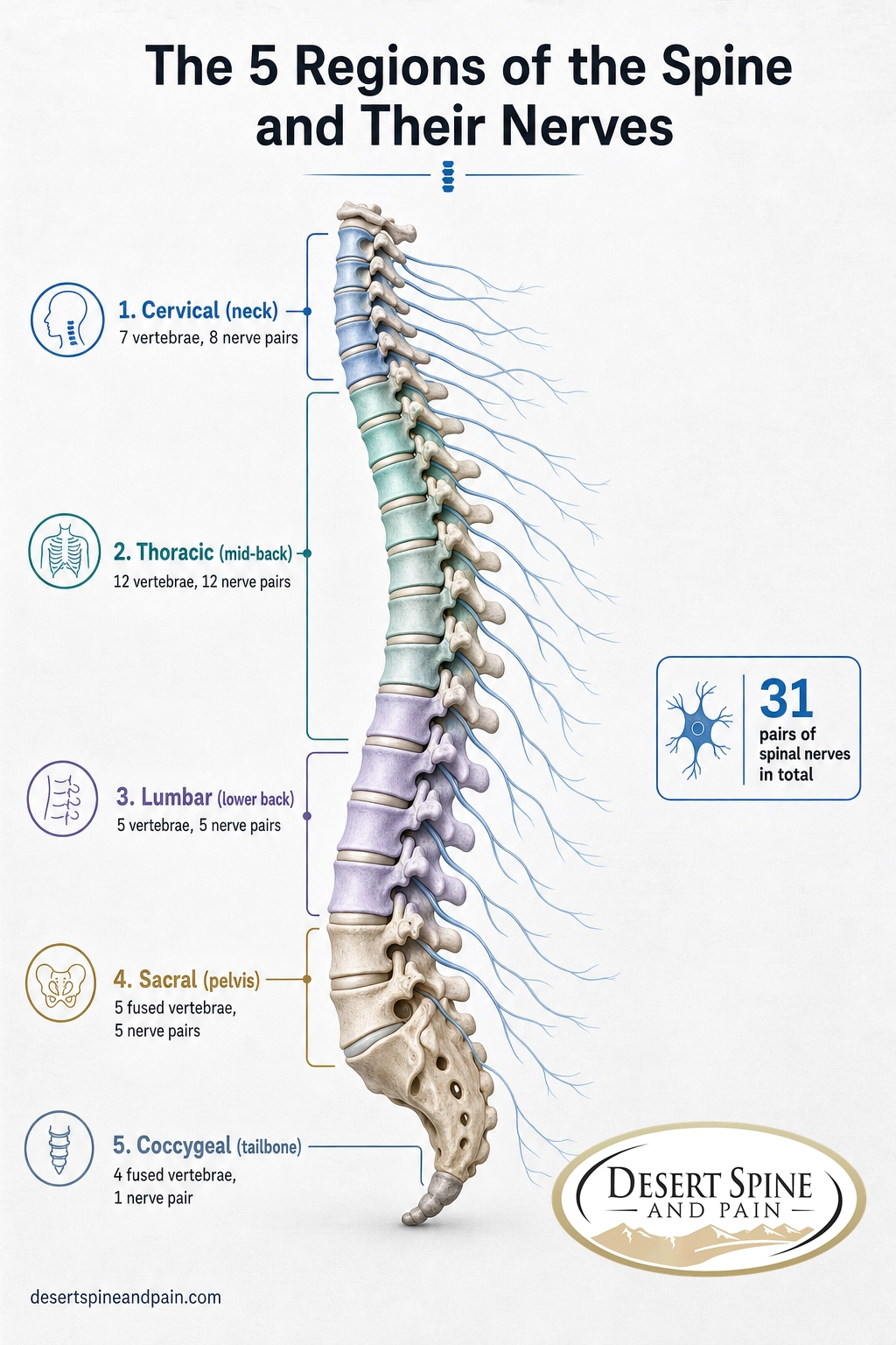
There are 31 pairs in total, grouped by region: 8 cervical (neck), 12 thoracic (mid-back), 5 lumbar (lower back), 5 sacral (pelvis), and 1 coccygeal (tailbone). The spine itself has 33 vertebrae, with 7 in the neck, 12 in the mid-back, 5 in the lower back, 5 fused into the sacrum, and 4 fused into the coccyx.
One detail trips up almost everyone, and it is worth getting straight before the chart, because it is the reason nerve numbers and vertebra numbers do not always match. In the neck, each of the first seven cervical nerves exits above its matching vertebra. That leaves room for an eighth cervical nerve, C8, which exits between the C7 and T1 vertebrae even though there is no eighth cervical vertebra. From T1 down through the rest of the spine, the pattern flips: each nerve exits below the vertebra of the same number. So the neck has 8 nerves for 7 bones, and everything below counts cleanly.
There is a second quirk lower down. The spinal cord does not run the full length of the spine. In most adults it tapers and ends around the first lumbar vertebra (L1) at a point called the conus medullaris. Below that, the canal is filled by a bundle of loose nerve roots, named the cauda equina (Latin for "horse's tail"), that keep traveling down before exiting at their own levels. That is why a lower-back problem can affect several leg and pelvic nerves at once.
Source: American Association of Neurological Surgeons: Anatomy of the Spine | StatPearls (NCBI): Neuroanatomy, Spinal Nerves
The Master Spinal Nerve Chart
This is the quick-reference map most people come here for. It pairs each spinal level with the main areas that level serves and the symptoms that typically appear when a nerve root at that level is compressed or irritated. Read it as a guide to where symptoms point, not as a diagnosis: nerve territories overlap from one person to the next, and only an in-person exam and imaging can confirm the level.
| Spinal level | Main areas it serves | Common symptoms when compressed |
|---|---|---|
| C1 to C4 | Back of the head, neck, upper shoulders; C3 to C5 feed the diaphragm through the phrenic nerve | Neck pain, headaches at the base of the skull, neck stiffness; high-level injury can affect breathing |
| C5 | Shoulder (deltoid), upper arm, biceps | Shoulder and upper-arm pain, weak shoulder lifting |
| C6 | Thumb side of the forearm and hand, wrist extensors | Pain or tingling into the thumb and index finger, weak wrist extension |
| C7 | Back of the arm, triceps, middle finger | Pain down the back of the arm, weak elbow straightening, numb middle finger |
| C8 | Little-finger side of the hand, finger flexors | Numbness in the ring and little fingers, weak grip |
| T1 | Inner forearm and elbow, small hand muscles | Inner-arm numbness, weak finger spreading |
| T2 to T12 | Chest wall and abdominal wall in horizontal bands (T4 at the nipple line, T10 at the navel) | Band-like pain wrapping around the chest or abdomen (uncommon; nerve roots here are rarely pinched) |
| L1 to L3 | Groin, front of the thigh, hip flexors, knee extension | Groin or front-thigh pain, weak hip flexion or knee straightening |
| L4 | Inner lower leg and ankle, ankle dorsiflexion, knee-jerk reflex | Inner-leg pain, weak lifting of the foot, reduced knee reflex |
| L5 | Outer lower leg, top of the foot, big toe | Pain down the outer leg to the top of the foot, weak big-toe lift, foot drop (a classic sciatica level) |
| S1 | Back of the leg, outer foot, heel, ankle plantarflexion | Pain down the back of the leg to the outer foot and heel, weak push-off, reduced ankle reflex |
| S2 to S5 | Back of the thigh, and bladder, bowel, and sexual function; the saddle area | Numbness in the saddle area, bladder or bowel changes (a red flag; see below) |
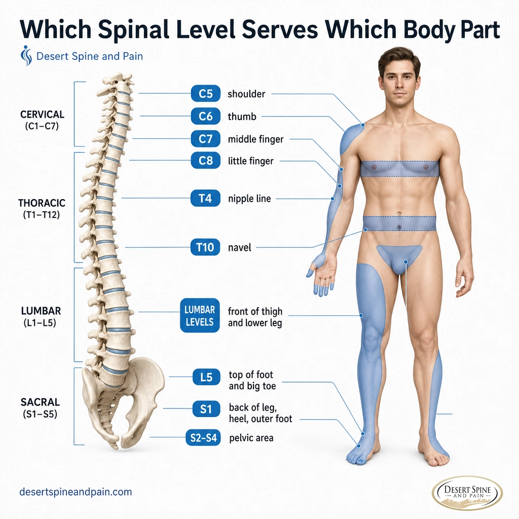
The sections below unpack each region, because the "why" behind the map is what makes it useful when symptoms do not fit neatly into one row.
Source: American Association of Neurological Surgeons: Anatomy of the Spine | StatPearls (NCBI): Anatomy, Skin, Dermatomes
Cervical Spine (C1 to C8): Neck, Arms, and Breathing
The neck is the busiest nerve hub in the spine. Its eight nerve pairs run everything from the muscles that hold your head up to the fine control in your fingertips, and they include one job no other region shares: breathing. The phrenic nerve, built mainly from the C3, C4, and C5 roots, drives the diaphragm, which is why high cervical injuries are the ones that threaten breathing.
For day-to-day nerve pinches, the lower neck matters most. A herniated disc or bone spur in the lower cervical spine irritates a specific root and produces a recognizable pattern: C5 pain tends to sit in the shoulder, C6 travels to the thumb, C7 runs down the back of the arm to the middle finger, and C8 reaches the little finger. Because these patterns are so consistent, a careful description of where your arm symptoms land often points to the level before any scan.
When a cervical root stays compressed, the symptom is called cervical radiculopathy, the medical term for a pinched nerve in the neck. If you are trying to sort out who to see about arm symptoms, our guide to which doctor treats a pinched nerve in the neck walks through the options. The underlying cause is often a disc problem, and you can read how an early tear in the disc wall sets the stage in our explainer on how an annular tear differs from a herniated disc.
Source: StatPearls (NCBI): Neuroanatomy, Spinal Nerves | Cleveland Clinic: Dermatomes
Thoracic Spine (T1 to T12): Chest and Trunk
The twelve thoracic nerves are the quiet workers of the spine. Each one wraps horizontally around the trunk as an intercostal nerve, supplying a band of skin and the muscles between the ribs and across the abdominal wall. Two landmarks make this region easy to picture: the T4 level sits at the nipple line, and the T10 level sits at the navel. If you ever feel a band of tingling or pain that circles part of your torso, that distribution is thoracic.
Because the thoracic spine is braced by the rib cage, it moves less than the neck or lower back, so disc herniations and pinched roots here are uncommon compared with the cervical and lumbar levels. When thoracic nerve symptoms do appear, they tend to show up as that band-like, wrap-around chest or abdominal discomfort rather than arm or leg pain, and they deserve a careful workup because chest and trunk symptoms have many possible causes.
A note on chest and trunk pain: band-like pain around the chest can come from a thoracic nerve, but chest pain can also signal heart or lung problems. New, severe, or unexplained chest pain is not something to map on a nerve chart at home. Seek prompt medical care to rule out urgent causes first.

Lumbar Spine (L1 to L5): Lower Back and Legs
The lower back carries the most mechanical load in the spine, and its five nerve pairs supply the hips and the fronts and sides of the legs. This is where a huge share of pinched-nerve symptoms begin, because the lumbar discs take the brunt of bending and lifting.
Moving down the region, the territory marches down the leg. The upper lumbar roots (L1 to L3) cover the groin and front of the thigh and power hip flexion and knee straightening. L4 covers the inner lower leg and helps lift the foot. L5, one of the two most common sciatica levels, sends symptoms down the outer leg to the top of the foot and the big toe, and a significant L5 pinch can weaken lifting the big toe or the whole foot, producing what clinicians call foot drop.
Remember the anatomy quirk from earlier: because the spinal cord ends around L1, the nerves traveling through the lower lumbar canal are already loose roots in the cauda equina. That means a single large disc herniation low in the back can press on more than one nerve at a time, which is why lumbar symptoms sometimes span several levels. When a lumbar root problem does not settle with time, targeted options range from an epidural steroid injection that calms inflammation around the root, to a foraminotomy that widens the opening where the nerve exits when a bone spur or disc is crowding it.
Source: American Association of Neurological Surgeons: Anatomy of the Spine | StatPearls (NCBI): Anatomy, Skin, Dermatomes
Sacral Spine (S1 to S5) and Coccyx: Pelvis and Feet
The sacrum is the broad, triangular bone at the base of the spine, formed from five fused vertebrae, and its five sacral nerve pairs exit through small openings in it. These nerves handle two very different jobs: the back of the legs and feet, and the pelvic organs.
On the movement side, S1 is the other classic sciatica level. It runs down the back of the leg to the outer foot and heel and powers push-off, so an S1 pinch can weaken standing on your toes and dampen the ankle reflex. The remaining sacral roots, S2 through S4, are where the nerve map turns critical: together they control the bladder, the bowel, and sexual function, and they supply sensation to the saddle area, the parts of the body that would touch a saddle. The single coccygeal nerve supplies a small patch of skin over the tailbone.
Because of what the sacral nerves control, their symptoms carry the most weight in this entire guide. Leg pain is miserable but rarely urgent; a new loss of bladder or bowel control is urgent. We will come back to that in the red-flags section.
Source: American Association of Neurological Surgeons: Cauda Equina Syndrome
Dermatomes and Myotomes: How Doctors Find the Level
Clinicians do not guess which level is involved. They lean on two overlapping body maps that turn your symptoms into a level, often before a scan confirms it.
Dermatomes (where you feel it)
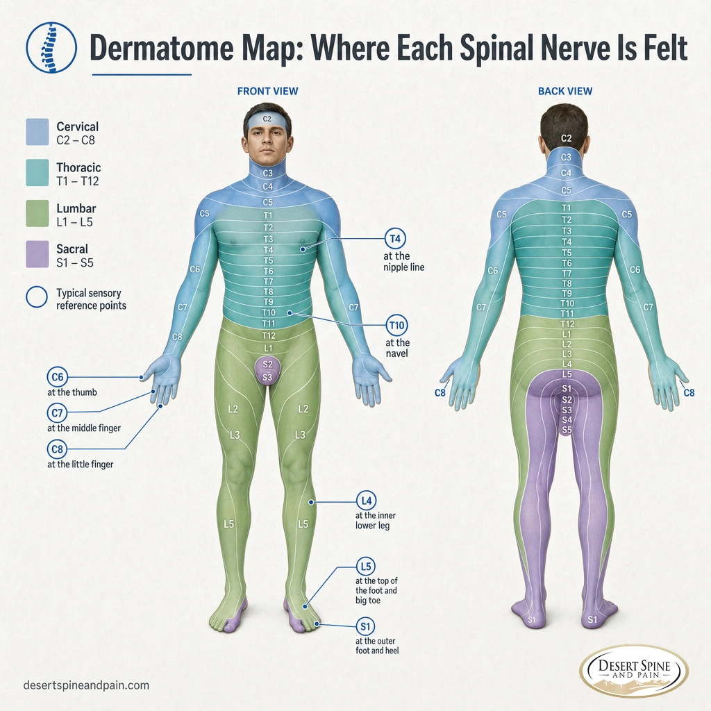
A dermatome is the patch of skin whose sensation is carried by a single spinal nerve. Map the exact path of your numbness or tingling and you are reading a dermatome backward to its nerve root. The map starts at C2, because the C1 nerve usually has no sensory branch, and the patches overlap with their neighbors, so the borders are approximate rather than exact. These are the landmarks clinicians lean on most:
| Nerve root | Skin landmark (dermatome) |
|---|---|
| C6 | Thumb |
| C7 | Middle finger |
| C8 | Little finger |
| T4 | Nipple line |
| T10 | Navel (umbilicus) |
| L4 | Inner lower leg and inner ankle |
| L5 | Top of the foot and the big toe |
| S1 | Outer foot and heel |
Myotomes (what gets weak)
A myotome is the group of muscles driven by a single spinal nerve root. Where dermatomes test feeling, myotomes test strength, and doctors use a short set of movements as a shortcut to each level. Most muscles draw on more than one root, so these are the signature tests, not the whole story:
| Nerve root | Signature movement (myotome) |
|---|---|
| C5 | Lifting the shoulder out to the side |
| C6 | Bending the wrist back (extension) |
| C7 | Straightening the elbow |
| C8 | Bending the fingers / gripping |
| T1 | Spreading the fingers apart |
| L2 | Bending the hip up |
| L3 | Straightening the knee |
| L4 | Pulling the foot up (dorsiflexion) |
| L5 | Lifting the big toe |
| S1 | Pushing the foot down / standing on tiptoe |
Put the two maps together and the logic is powerful. Numbness in the little finger (a C8 dermatome) plus a weak grip (a C8 myotome) points firmly at the C8 root. Tingling on the top of the foot (L5) plus a weak big-toe lift (L5) points at L5. This is exactly the reasoning a spine specialist uses, then confirms with an MRI and, when needed, nerve testing such as EMG.
Source: Cleveland Clinic: Dermatomes | StatPearls (NCBI): Anatomy, Skin, Dermatomes
From Symptom to Level: A Quick Decision Guide
Here is how to read your own symptoms against the map. Think of this as a way to have a sharper conversation with your doctor, never as a substitute for an exam.
- Symptoms in the shoulder or down the arm to the hand: suspect a cervical level (C5 to T1). Where it lands in the hand narrows it: thumb points to C6, middle finger to C7, little finger to C8.
- A band of pain or tingling wrapping around the chest or abdomen: suspect a thoracic level (and rule out non-spinal causes first).
- Pain or tingling down the leg to the top of the foot and big toe, with a weak big-toe lift: suspect L5.
- Pain down the back of the leg to the outer foot and heel, with weak push-off: suspect S1.
- Groin or front-of-thigh symptoms with weak knee straightening: suspect an upper lumbar level (L2 to L4).
- Numbness in the saddle area or new bladder or bowel changes: treat as an emergency, not a chart entry. See the next section.
The reason this works is the same reason the whole guide works: the spine is wired in an orderly way, so symptoms are a return address. The catch is that an MRI often shows age-related changes at several levels, and not every finding is the one causing your pain. Matching the symptom pattern to a specific dermatome and myotome is how a specialist separates the guilty level from the incidental ones, which is why a hands-on exam still beats a scan read in isolation. Our overview of common spine conditions and how they are evaluated covers what that workup looks like.
Red Flags: When a Pinched Nerve Is an Emergency
Most pinched nerves are painful but not dangerous, and the large majority improve with time and non-surgical care. A small set of symptoms are the exception, and they come from the two parts of the system that cannot wait: the spinal cord and the lowest nerve roots.
Seek immediate medical care if you have any of these:
- Saddle numbness (loss of feeling in the inner thighs, groin, or buttocks).
- New loss of bladder or bowel control, including urinary retention (being unable to go) or incontinence.
- Weakness or numbness in both legs, especially if it is worsening.
- In the neck: clumsy hands, dropping objects, or new trouble with balance and walking.
The first three can signal cauda equina syndrome, in which the bundle of lower nerve roots is compressed, most often by a large lumbar disc herniation. It is a surgical emergency: specialists aim to relieve the pressure urgently, and treatment within about 48 hours of onset offers a meaningful advantage, because delays risk permanent loss of bladder, bowel, and leg function. The neck cluster can signal cervical myelopathy, pressure on the spinal cord itself rather than a single root, which also needs prompt evaluation. If any of these describe you, do not wait to map a dermatome; get care now.
Source: American Association of Neurological Surgeons: Cauda Equina Syndrome
Why One Expert Across the Whole Spine Matters
Reading the nerve map is the easy part. Acting on it is where experience counts, because the same symptom can come from a disc, a bone spur, narrowing of the canal, or a problem that is not in the spine at all. The clinician who can sort that out is one who understands both the nerves and the bones that surround them, and who can carry you from the first exam through conservative care and, only if you ever need it, surgery.
At Desert Spine and Pain, that range sits with one surgeon. The practice is led by Dr. David L. Greenwald, MD, FACS, who is board certified as both a spine surgeon and a neurosurgeon. That dual training covers the whole picture mapped in this guide, from the nerve root and the spinal cord to the vertebrae and discs pressing on them, so a single expert can pinpoint the level, explain what it means, and choose the least invasive option that fits. The practice leads with non-surgical care and reserves surgery for when the evidence genuinely calls for it.
Desert Spine and Pain serves the Greater Phoenix, Arizona area, welcomes out-of-network patients, and partners with personal injury attorneys and their clients. If pain, numbness, or weakness is tracing one of the patterns above, a focused consultation is the fastest way to find out which level is involved and what to do about it.
Meet Dr. David L. Greenwald, MD, FACS
Explore treatments for nerve compression
Book a consultation or second opinion
Common Questions About Spinal Nerves
Which vertebrae affect which nerves?
How many spinal nerves does the human spine have?
Why are there 8 cervical nerves but only 7 cervical vertebrae?
Which nerve causes sciatica down the leg?
What symptoms mean a pinched nerve is an emergency?
Related Spine Terms
- Spinal nerve root: the part of a spinal nerve closest to the cord, where compression usually happens.
- Dermatome: the patch of skin whose sensation is carried by a single spinal nerve.
- Myotome: the muscle group driven by a single spinal nerve root.
- Radiculopathy: the medical term for a pinched or irritated nerve root, named by region (such as cervical or lumbar).
- Conus medullaris: the tapered lower end of the spinal cord, usually around the L1 vertebra.
- Cauda equina: the bundle of loose nerve roots below the cord's end; compression here is an emergency.
- Annular tear: a tear in the outer wall of a disc that can precede the herniation behind many pinched nerves.
Sources
Sources and Methodology
This guide summarizes established clinical anatomy from peer-reviewed references and major medical institutions. Nerve territories, dermatome landmarks, and myotome tests are standard teaching maps: they overlap between neighboring levels and vary modestly from person to person and between textbooks, so they are presented for orientation, not as a diagnosis. This is educational information, not medical advice. Any new or worsening pain, numbness, or weakness should be evaluated by a qualified physician in person, and the emergency symptoms described above need immediate care.
- American Association of Neurological Surgeons (AANS): Anatomy of the Spine and Peripheral Nervous System
- American Association of Neurological Surgeons (AANS): Cauda Equina Syndrome
- StatPearls (NCBI Bookshelf): Neuroanatomy, Spinal Nerves
- StatPearls (NCBI Bookshelf): Anatomy, Skin, Dermatomes
- Cleveland Clinic: Dermatomes